Echo Cardiography
Echo Cardiography
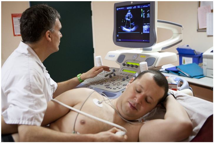
DescriptionDuring an echocardiogram, sound waves are used to produce images of your heart. With this typical test, your doctor can watch your heart beating and pumping blood. Your doctor can use it to determine whether you have heart disease.
Your echo is performed by a technician known as a cardiac sonographer. Dr. Priyanka Rana of Param Imaging train to perform echo tests and uses cutting edge technology. She trained to work in different environments, including hospital
rooms and catheterization labs.
An echocardiogram and an electrocardiogram (also known as an EKG or ECG) examine your heart. However, they check for different things and generate different kinds of visuals. An echocardiogram examines the overall structure and function of your heart. It creates moving images of your heart.
An EKG examines the electrical activity of your heart. Instead of images of your heart, it generates a graph. The lines on this graph represent your heart rate and rhythm.
Why is an echocardiogram done in a pregnancy?
Fetal echocardiography is a type of ultrasound test. This exam allows your doctor to see the structure and function of your unborn child’s heart in greater detail.
A fetal echocardiogram is not for all pregnant women. A basic ultrasound will show the development of all four chambers of the baby’s heart for the majority of women. If previous tests were inconclusive or detect an abnormal heartbeat in the fetus, your OB-GYN may recommend this procedure.
The results of this test will assist you and Dr. Priyanka Rana in planning any post-delivery treatments your baby may require. You can also get help and counseling to make good decisions for the rest of your pregnancy.